
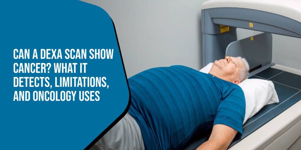
When you or a loved one undergo health screenings, it is natural to hope that a single test might catch multiple underlying medical conditions. If you have been scheduled for a dual-energy X-ray absorptiometry scan, you might reasonably wonder: can a DEXA scan show cancer during a routine evaluation? The short answer is no—a DEXA scan is not designed or calibrated as a cancer detection tool. However, the full clinical picture is far more nuanced.
While a DEXA scan primarily quantifies bone mineral density (BMD) and body composition, subtle bone changes caused by advanced malignancies can occasionally catch an radiologist’s eye. Understanding what a DEXA scan actually measures—and recognizing its limitations—helps prevent misplaced reliance on a test intended for bone density rather than oncology diagnostics.
In this comprehensive guide, we examine what DEXA technology can and cannot detect, how cancer treatments intersect with bone health, and which specialized imaging modalities medical professionals rely on for accurate cancer screening and diagnosis.
Can a DEXA scan show cancer?
No, a standard DEXA scan cannot reliably detect or diagnose cancer. DEXA scans use low-dose X-rays to measure bone mineral density (BMD) and body composition (fat mass and lean muscle mass). They lack the structural resolution and metabolic soft-tissue sensitivity needed to spot early-stage tumors or cancers.
- Primary Function: Diagnosing osteoporosis, osteopenia, and evaluating body composition.
- Can It Detect Tumors? Only in rare instances where a large tumor or aggressive bone metastasis significantly destroys local bone density (lytic lesions).
- Best Scans for Cancer: MRI, CT scans, PET scans, Mammograms, and Bone Scans.
What Is a DEXA Scan and How Does It Work?
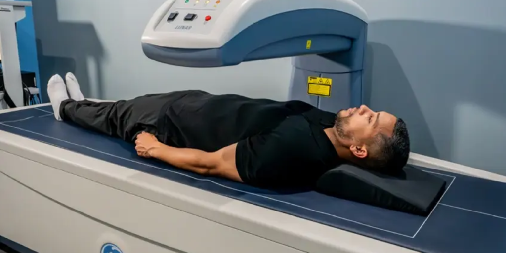
A dual-energy X-ray absorptiometry (DEXA or DXA) scan is a non-invasive, low-dose imaging technique considered the clinical gold standard for measuring bone density. During a typical 10-to-15-minute examination, two distinct low-energy X-ray beams pass through targeted body regions—most commonly the hip, lumbar spine, and forearm.
Because dense bone tissue absorbs X-ray photons differently than soft tissue (muscle and fat), sophisticated detector software calculates precisely how much mineral density is present. The results are reported primarily as a T-score (comparing your bone density to a healthy young adult) and a Z-score (comparing your bone density to peers of your age, sex, and ethnicity).
Key Metrics Delivered by DEXA Technology
- Bone Mineral Density (BMD): Evaluates structural bone strength to identify osteoporosis or osteopenia.
- Visceral Adipose Tissue (VAT): Measures deep belly fat surrounding internal organs, a key marker for metabolic health.
- Lean Muscle Mass: Quantifies skeletal muscle percentage to monitor physical training, sarcopenia, or athletic recovery.
- Total Body Fat Percentage: Delivers precise body composition breakdowns superior to basic BMI or caliper assessments.
Why the Question Matters: DEXA Scan Cancer Detection vs. Diagnosis
Understanding whether a DEXA scan detects cancer is vital for two primary reasons: avoiding false security and ensuring proper medical follow-up. Believing that a clear bone density report equates to a clean bill of health regarding cancer can cause individuals to miss routine screenings like mammograms, colonoscopies, or low-dose CT scans.
Furthermore, many cancer patients are explicitly ordered to get DEXA scans by their oncologists. This frequent overlap leads patients to assume the scan is searching for cancer recurrence, when in reality, it is monitoring collateral bone loss from cancer therapies.
Can a DEXA Scan Detect Cancer or Bone Tumors?
To directly answer whether a DEXA scan can detect tumors or malignancies: it is extremely rare and clinically unreliable.
DEXA machines operate on extremely low radiation levels—roughly 1/10th to 1/20th the radiation of a standard chest X-ray. While this makes DEXA exceptionally safe for routine health tracking, it drastically limits soft-tissue visualization. Tumors originating in soft tissues (such as breast, lung, colon, or prostate tissue) remain entirely invisible on a DEXA scan.
Can a DEXA Scan Show Bone Cancer or Metastasis?
Bone cancer—either primary bone sarcomas or metastatic cancer that has spread from organs to the skeleton—primarily affects bone architecture in two ways:
- Lytic Lesions: Cancer cells eat away at bone tissue, creating localized holes or weak spots.
- Blastic Lesions: Cancer cells stimulate abnormal, disorganized bone overgrowth.
If a lytic lesion is exceptionally large and located directly within the regional region scanned (such as the femoral neck or lumbar vertebrae), a DEXA reading might show an unexpected, drastic drop in bone density in that isolated spot. A radiologist reading the scan may flag this anomaly as a potential structural defect requiring secondary evaluation. However, DEXA cannot differentiate whether that density loss stems from severe focal osteoporosis, a benign bone cyst, an infection, or a malignant bone tumor.
Comparing Imaging Modalities: DEXA vs. Cancer Detection Scans
Different imaging technologies serve distinct diagnostic roles. The table below illustrates how DEXA compares against diagnostic scans explicitly designed for cancer detection:
| Imaging Modality | Primary Diagnostic Target | Soft Tissue Resolution | Radiation Level | Effective for Cancer Detection? |
|---|---|---|---|---|
| DEXA Scan | Bone mineral density, body composition | Very Low | Micro-dose (<0.001 mSv) | No (Only detects severe local bone loss) |
| MRI (Magnetic Resonance) | Soft tissue, brain, spinal cord, joints | Exceptional | None (Magnetic fields) | Yes (High detail for tumors & lesions) |
| CT Scan (Computed Tomography) | Cross-sectional organ and tissue detail | High | Moderate to High | Yes (Locates tumor size, shape, spread) |
| PET Scan (Positron Emission) | Cellular metabolic activity | Moderate (Paired with CT/MRI) | Moderate (Radioactive tracer) | Yes (Identifies active, growing cancer cells) |
| Nuclear Bone Scan | Whole-body bone turnover & metabolic activity | Low (Focuses on bone) | Moderate (Tracer absorbed by bone) | Yes (Standard for detecting bone metastases) |
The Crucial Link Between Cancer Treatments and DEXA Scans
Although DEXA scans do not screen for cancer, they are an essential tool in comprehensive cancer care management. Many life-saving oncological treatments severely impact bone metabolism, placing patients at elevated risk for rapid bone loss, osteopenia, fragile fractures, and severe osteoporosis.
1. Hormone Therapy for Breast and Prostate Cancer
Estrogen and testosterone play crucial roles in maintaining bone density. Certain cancer treatments actively suppress these hormones to slow tumor growth:
- Aromatase Inhibitors (AIs): Used in hormone-receptor-positive breast cancer, AIs drastically lower estrogen levels, often causing accelerated bone loss.
- Androgen Deprivation Therapy (ADT): Used in prostate cancer care, ADT reduces testosterone, which significantly lowers bone mineral density over time.
2. Chemotherapy and Radiation
Chemotherapy agents can damage osteoblasts (bone-building cells) and induce premature menopause in premenopausal women, triggering sharp drops in density. Similarly, direct radiation therapy to areas like the pelvis can weaken local bone structure.
3. Corticosteroid Use
High-dose glucocorticoids (like dexamethasone or prednisone) are routinely prescribed alongside chemotherapy to curb nausea and allergic reactions. Prolonged corticosteroid exposure is a leading cause of drug-induced osteoporosis.
In all these scenarios, baseline and follow-up DEXA scans allow care teams to introduce bone-protective medications (such as bisphosphonates or RANKL inhibitors) before a painful fracture occurs.
Common Misconceptions About DEXA Scans and Cancer
Misconception 1: “My DEXA scan was clear, so I don’t need a mammogram or CT.”
Fact: A DEXA scan does not evaluate soft tissues where most primary cancers develop. Regular screening mammograms, Pap smears, colonoscopies, and low-dose lung CTs remain indispensable regardless of DEXA results.
Misconception 2: “A low T-score on my DEXA means I have bone cancer.”
Fact: Low bone density (a low T-score) is overwhelmingly caused by age-related osteoporosis, dietary calcium/vitamin D deficiencies, hormonal changes, or lack of weight-bearing exercise. Bone cancer is a rare cause of low T-scores.
Misconception 3: “DEXA scans use dangerous amounts of radiation that cause cancer.”
Fact: A standard body composition or bone density DEXA scan exposes you to less background radiation than a typical cross-country flight from Boston to San Francisco. The cancer risk from a DEXA scan is virtually non-existent.
Best Practices: When to Request Diagnostic Cancer Imaging
If you are experiencing unexplained physical symptoms, relying on a bone density test will not give you clear answers. Medical guidelines recommend contacting a primary care physician to request dedicated diagnostic workups if you observe any of the following signs:
- Unexplained, rapid weight loss without changes in diet or exercise.
- Persistent, localized bone pain that worsens at night or does not improve with rest.
- Unexplained fatigue, persistent low-grade fever, or night sweats.
- New, unusual lumps or swelling in soft tissue, lymph nodes, or joints.
- A personal history of cancer combined with sudden back pain, hip pain, or joint discomfort.
Step-by-Step: What to Expect During a DEXA Scan
If your doctor orders a DEXA scan to check your bone health or if you are using it to track lean muscle mass and fat distribution, the process is quick, painless, and simple.
- Preparation: Wear comfortable, loose clothing without metal zippers, snaps, or buttons. Avoid taking calcium supplements for at least 24 hours prior to the test.
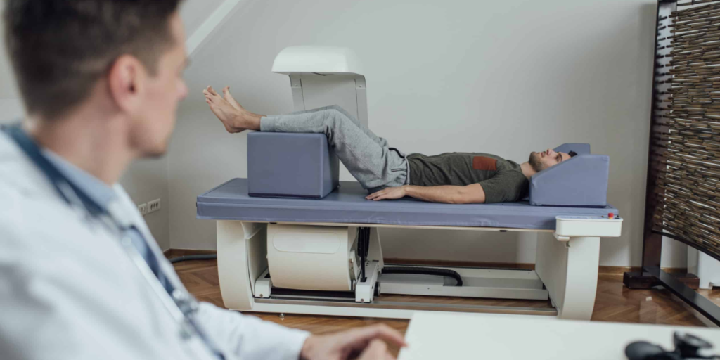
- Positioning: You lie flat on a padded open table. The technologist positions your legs or arms using soft foam blocks to align your hip and spine correctly.
- The Scan: A motorized arm passes slowly over your body. Unlike an MRI, the machine is completely open, quiet, and non-claustrophobic.
- Duration: The actual imaging takes between 5 and 15 minutes.
- Results: A certified radiologist analyzes the images and provides a report to your physician, who will review your T-score, Z-score, or body composition breakdowns with you.
Proactive Health Management: Integrating Bone Density & Fitness
Maintaining optimal health requires an integrated approach that pairs medical diagnostics with targeted lifestyle interventions. Whether you are managing post-treatment recovery or simply taking charge of your long-term wellness, optimizing bone mineral density and body composition requires structured resistance training.
High-impact, progressive resistance exercises signal bone-building cells to deposit new mineral matrices, strengthening bones while simultaneously increasing lean muscle mass and improving metabolic health.
For independent personal trainers and clients in the Boston area seeking an elevated, distraction-free environment to conduct customized strength and conditioning programs, Back Bay Fit provides state-of-the-art training facilities designed for personalized performance, rehabilitation support, and long-term vitality.
Frequently Asked Questions (FAQs)
Can a DEXA scan show bone cancer?
A DEXA scan cannot definitively show or diagnose bone cancer. While severe, localized bone destruction caused by advanced tumors might appear as an unexplained, drastic dip in bone density on a DEXA image, the scan lacks the sensitivity to identify cancer. Suspected bone cancer requires dedicated nuclear bone scans, MRIs, CT scans, or tissue biopsies for confirmation.
What can a DEXA scan detect?
A DEXA scan primarily detects osteopenia (low bone mass), osteoporosis (severe bone loss), and overall bone fracture risk. When used for body composition analysis, it precisely measures body fat percentage, lean muscle mass distribution, and visceral adipose tissue (VAT).
Can a DEXA scan detect tumors?
No, DEXA scans cannot detect soft-tissue tumors or early-stage bone tumors. The low-dose X-ray beams used in DEXA technology are optimized specifically for density calculations across bone and mass tissue, not for rendering high-resolution details of soft tissues or cellular masses.
What scan is best for detecting cancer?
The best scan depends entirely on the suspected type and location of the cancer. MRI scans excel at evaluating soft tissues, the brain, and spinal structures. CT scans provide detailed cross-sectional views of chest, abdominal, and pelvic organs. PET scans highlight active metabolic cancer cells throughout the body, and Nuclear Bone Scans are ideal for detecting bone metastases.
Conclusion
To summarize, if you are asking can a DEXA scan show cancer, the clinical reality is clear: DEXA scans are engineered for evaluating bone density and body composition, not for detecting, staging, or diagnosing malignant tumors. While a DEXA scan plays a vital supportive role in monitoring bone health during cancer therapies, it should never replace dedicated screening tools such as MRIs, CT scans, mammograms, or PET imaging.
By understanding the precise function of DEXA technology, you can make informed choices about your healthcare, maintain essential cancer screenings, and take proactive steps to protect your bone strength and physical fitness through every stage of life.
